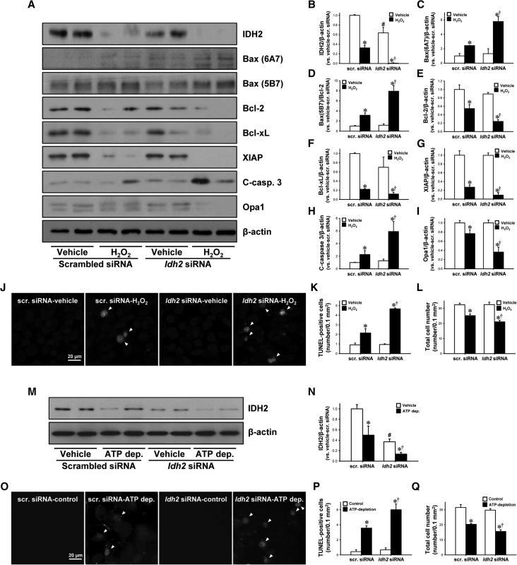
Figure 8.
Effect of IDH2 downregulation on H2O2-induced apoptosis in cultured mProx24 cells. mProx24 cells were transfected with Idh2-siRNA and scrambled siRNA (Scr-siRNA). After transfection, cells were treated with 500 µM H2O2 for 5 hours. (A) Cell lysates were analyzed by Western blotting using antibodies directed against IDH2, Bax (6A7), Bax (5B7), Bcl-2, Bcl-xL, XIAP, cleaved caspase-3 (C-casp-3), and Opa1. β-actin was used as a loading control. (B–I) The densities of bands were measured using ImageJ software. (J) Fixed cells were subjected to TUNEL assay. Arrows indicate TUNEL-positive cells. TUNEL-positive (K) and total cell number (L) were determined. (M–Q) mProx24 cells transfected with either Idh2-siRNA or Scr-siRNA were incubated in Krebs–Henseleit buffer with or without sodium cyanide (5 mM) and 2-deoxyglucose (5 mM) for 60 minutes, followed by Krebs–Henseleit buffer containing 10 mM of dextrose for 40 minutes. Cells were subjected to Western blot analysis using anti-IDH2 antibody. β-actin was used as a loading control. (N) The densities of bands were measured using ImageJ software. (O) Fixed cells were subjected to TUNEL assay. Arrows indicate TUNEL-positive cells. TUNEL-positive (P) and total cell number (Q) were counted. Results are expressed as mean±SEM (n=3). *P<0.05 versus respective vehicle-treated siRNA; †P<0.05 versus H2O2-treated or ATP-depleted Scr. siRNA; #P<0.05 versus vehicle-treated Scr. siRNA. ATP dep., ATP depletion.

