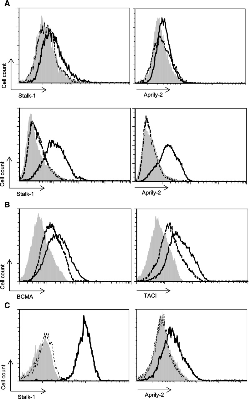
Figure 5.
TLR9 activation induces APRIL expression in tonsillar B cells. (A) Tonsillar B cells isolated from patients with CT were stimulated daily with 10 μg/ml CpG. APRIL expression is shown on viable (upper panel) and permeabilized (lower panel) gated CD19+ B cells. (B) Surface expressions of TACI and BCMA are also shown. (C) CD19+ B cells from patients with CT were purified on an FACS ARIA (BD Pharmingen) by positive selection. Purified CD19+ B cells were stimulated daily with 10 μg/ml CpG. APRIL expression is shown on permeabilized cells. Shaded histograms represent control isotype–matched reactivity. Dotted and straight lines represent indicated antibody reactivities on control and CpG-ODN–stimulated cells, respectively, at day 7. Histogram plots are representative of at least three experiments performed with tonsils from independent patients.

