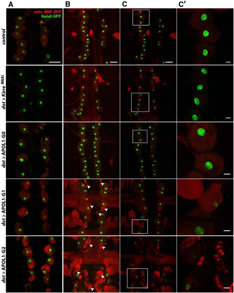
Figure 2.
APOL1 G1 and G2 affect Drosophila pericardial nephrocyte function and lead to cell death during aging. Pericardial nephrocytes of adult flies were dissected, and ANF-RFP uptake and nuclear GFP were analyzed by confocal microscopy. (A) ANF-RFP uptake by pericardial nephrocytes in 1-day-old flies. (B and C) ANF-RFP uptake by pericardial nephrocytes in 15-day-old flies. In B, the maximal intensity projection of 20 focal planes was applied to visualize fragments of dead cells (arrowheads). Close-up images are shown in C′ (corresponding to the white frames in C). Genotypes used in this experiments were (from top to bottom) with myosin heavy chain (MHC)-ANF-RFP, Hand-GFP, Dot-GAL4/+ (control); with MHC-ANF-RFP, Hand-GFP; Dot-GAL4/UAS-KirreRNAi (Dot > KirreRNAi); with MHC-ANF-RFP, Hand-GFP; Dot-GAL4/+;UAS-APOL1-G0 (Dot > APOL1-G0); with MHC-ANF-RFP, Hand-GFP; Dot-GAL4/+;UAS-APOL1-G1 (dot > APOL1-G1), and with MHC-ANF-RFP, Hand-GFP; Dot-GAL4/+; UAS-APOL1-G2 (dot > APOL1-G2). Scale bars, 50 μm in A–C; 10 μm in C′.

