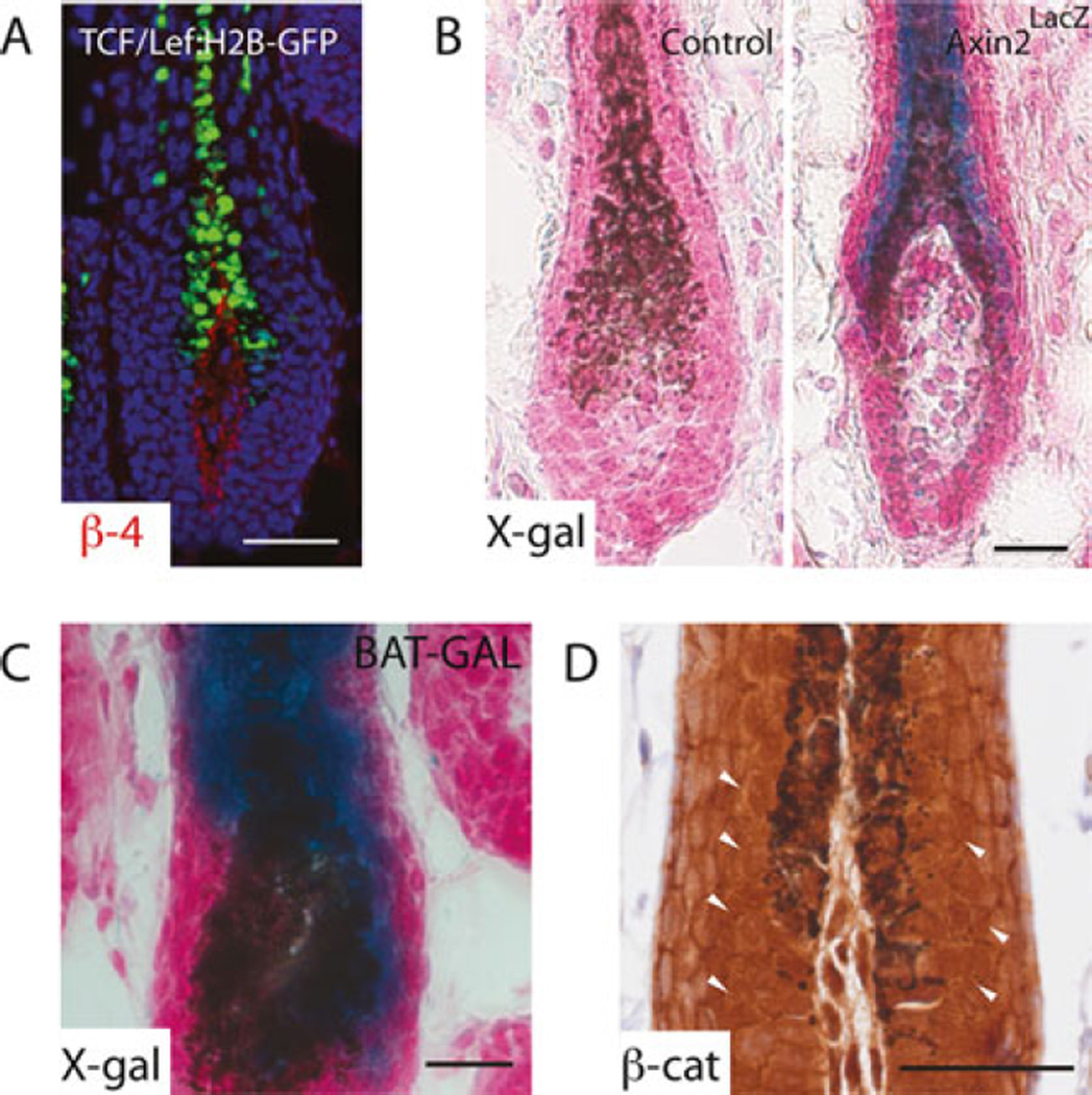
Fig. 3.
Detecting Wnt/β-catenin signaling. (a) Fluorescence image of endogenous GFP signal in the hair follicle of 28-day-old TCF /LEF:H2B-GFP mouse, with nuclei counterstained with Hoechst. β4-integrin co-staining in red labels the basement membrane, delineating the interface between epithelial cells and the dermal papilla. X-Gal staining of 28-day-old (b) wild-type or Axin2lacZ and (c) BAT-GAL mice. Skins were counterstained with Nuclear Fast Red to mark nuclei. Blue signal in the precortex of hair follicle indicates Wnt activity. (d) Immunostaining of β-catenin in 28-day-old wild-type mouse skin. Arrowheads point to nuclear β-catenin. Scale bar denotes 50 µm

