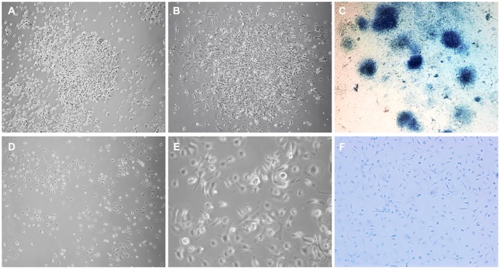
Fig 1. Comparison of morphological appearance of amniotic fluid cells from MMC and normal fetuses.
Representative phase-contrast micrographs of cells isolated from MMC-AF at E21 illustrate clusters of densely packed amniotic fluid cells (A) at 24 hours after plating, (B) at 72 hours after plating, and (C) image of distinct clusters of cells stained with methylene blue at 72 h after plating. In contrast, representative micrographs illustrate a loose monolayer growth pattern of amniotic fluid cells isolated from age-matched normal controls shown at (D) lower and (E) higher magnification, and (F) image of these cells stained with methylene blue at 72 h after plating showing the absence of cell clusters (compare F and C). (Magnification A, B, D, F10X; E20 X; C4X).

