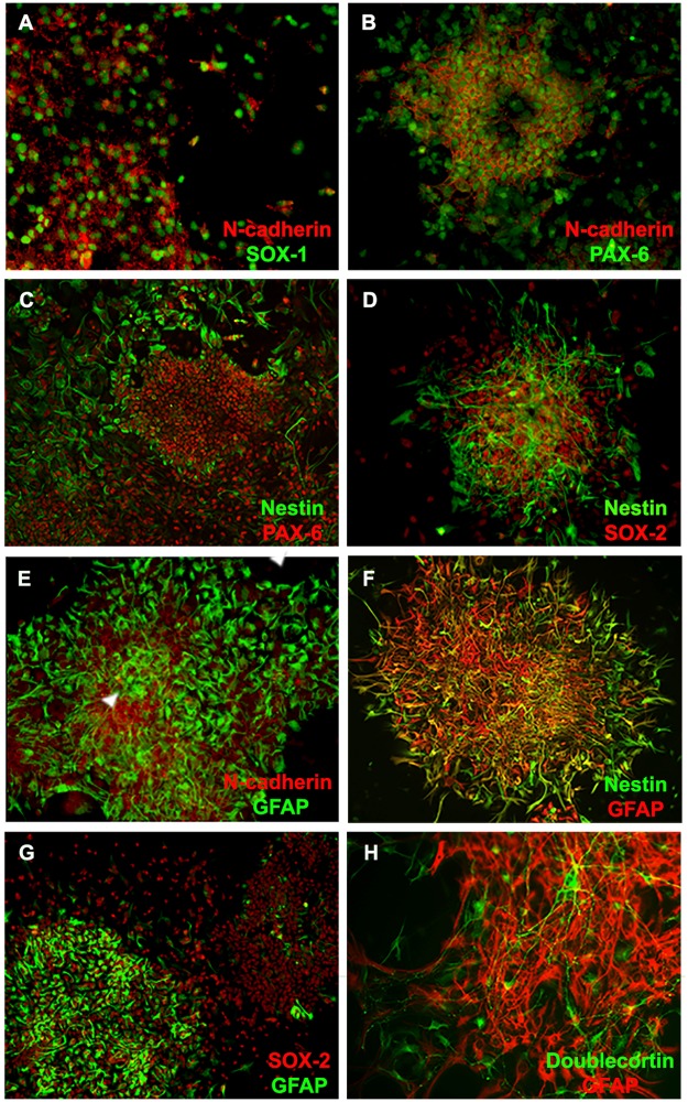
Fig 3. Expression of neural markers by amniotic fluid cells isolated from MMC.
(A-D) Double immunofluorescence staining demonstrates that the majority of cells within N-cadherin positive clusters stained positive for markers of early neuroepithelial cells such as SOX-1 and Pax-6 along with other progenitor markers such as nestin and SOX-2. (E and F) Double immunofluorescence staining for the expression of N-cadherin and GFAP show N-cadherin-positive clusters with areas of strong junctional expression of this protein (arrowhead) contained GFAP positive cells. (G) Double immunofluorescence staining for the expression of GFAP and Nestin shows GFAP positive cells, which often co-expressed nestin as wells as GFAP positive cells that were negative for nestin. (H) Double immunofluorescence staining for the expression of GFAP and SOX-2 demonstrates clusters with high and low fraction of GFAP positive cells. (I) GFAP positive cells within clusters were also accompanied by doublecortin positive neurons. (Magnification 20 X).

