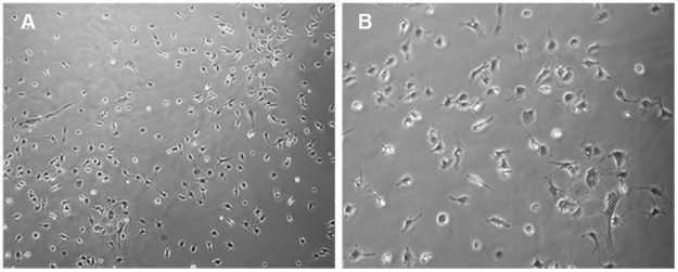
Fig 4. A monolayer of amniotic fluid cells from MMC fetuses at E19.
(A and B) At 72 h after plating, representative phase-contrast micrographs of amniotic fluid cells illustrate a monolayer growth pattern of amniotic fluid cells in the absence of cell clusters in cultures of amniotic fluid cells isolated from MMC-AF obtained at E19. (Magnification (A 10 X; B20 X).

