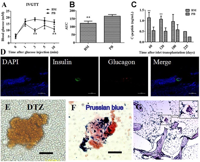
Fig 3. The identification of islets in the bone marrow cavity.
Comparison of blood glucose levels (Fig 3A) and AUC for glucose (Fig 3B) in the peripheral blood (PB) and bone marrow (BM) during IVGTT following islet transplantation in Group 3. The C-peptide level (Fig 3C) was measured in the bone marrow and peripheral blood. Tibial bone was harvested 225 days after transplantation and stained for insulin and glucagon to confirm islet survival. The bone marrow collected from the tibia 225 days after islet transplantation was stained with DTZ(Fig 3E) and Prussian blue (Fig 3F). Tibial bone was stained for insulin and Prussian blue to confirm SPIO (Fig 3G).**, P < 0.01 versus PB group.

