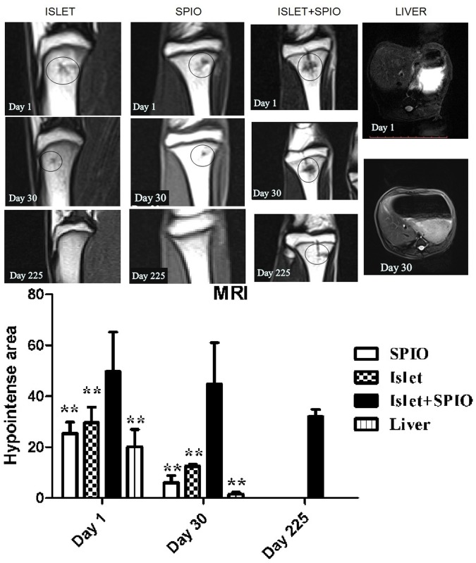
Fig 4. MRI of SPIO labeled islets in vivo.
MRI was performed 1, 30 and 225 days after islet(left), SPIO (left middle) and islet+SPIO (right middle) transplantation. MRIs of the liver were acquired 1and 30 days after islet+SPIO (right) transplantation. A small hypo-intense area (black circle) inside the normal hyperintense signal was evident at the site of the islet infusion at the level of the tibia. The area of hypointense spots visible in the bone marrow tissue and liver in each slice in different groups (below). **, P < 0.01 versus islet+SPIO group.

