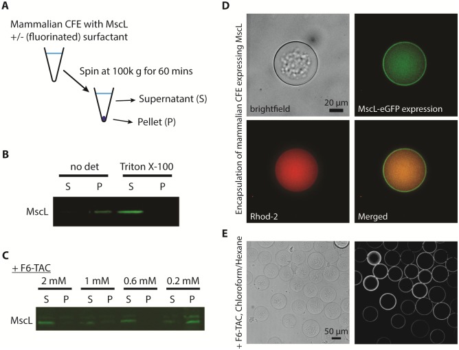
Fig 3. CFE of MscL is solubilized in fluorinated surfactants.
(A) Schematic illustration of the solubility assay for CFE of MscL. (B) Solubility of CFE of MscL after 5 hr in the presence or absence of 0.2% of Triton X-100. (C) Solubility of CFE of MscL after 5 hr at different concentrations of fluorinated surfactant F6-TAC. (D) Formation of vesicles encapsulating mammalian CFE expressing MscL in the presence of 2% PVA and 0.6 mM F8-TAC. The vesicles were formed from 36/64 chloroform/hexane as the middle phase, and imaged in brightfield (top left) and in green fluorescence (top right). 5 μM of calcium indicator Rhod-5N and 1 mM of calcium was also encapsulated inside the vesicle (bottom left). Merged image of green and red fluorescence is shown in the bottom right. (E) Formation of HeLa lysate encapsulated vesicles formed from 40/60 chloroform/hexane in the presence of 2% PVA and 2 mM F6-TAC, imaged in brightfield (left) and in fluorescence (right).

