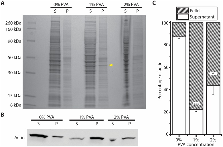
Fig 7. Actin aggregates with PVA surfactant.
(A) Coomassie blue stained gel showing proteins in the supernatant or pellet after 10 min centrifugation at 16,100 g, in the presence of PVA surfactant at different concentrations. (B) Western blot showing the actin in the supernatant or pellet fractions at different PVA concentrations. (C) Percentage of actin measured in the supernatant and pellet fractions at different PVA concentrations (n = 3, ±S.E.), unpaired t test comparing with 0% PVA; *: p < 0.01; ***: p < 0.0001.

