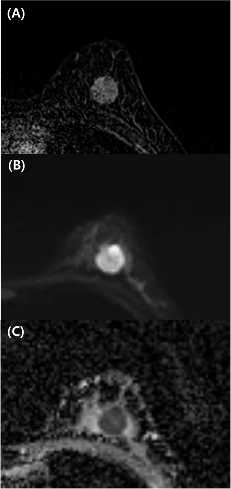
Fig 2. 33-year-old woman with invasive ductal carcinoma in the left breast.

Contrast-enhanced T1-weighted axial image (A), readout-segmented echo-planar DWI image (B), and ADC map (C). (A) Round circumscribed mass with heterogeneous enhancement in the breast. With DWI, at 750 seconds/mm2, there is a round circumscribed mass with heterogeneous high signal intensity in the left breast (B) with low ADC (0.8×10⁻³ mm2/sec) (C). The patient underwent breast-conserving surgery. The final diagnosis was invasive ductal carcinoma of histological grade III and triple-negative subtype.
