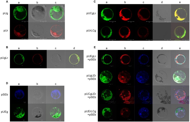
Fig 5. Protoplasts transformed by subcellular locational vectors.
(A) Protoplasts transformed by pUg and pUr. A: GFP excitation at 488 nm and RFP excitation at 543 nm; B: light field; and C: AB merge; scale bar = 10 μm. (B) Protoplasts transformed by pUgLr. A: GFP excitation at 488 nm; B: RFP excitation at 543 nm; C: light field; and D: ABC merge; scale bar = 10 μm. (C) Protoplasts transformed by pUCgLr and pUrLCg. A: GFP excitation at 488 nm; B: RFP excitation at 543 nm and emission at 550–710 nm; C: RFP excitation at 543 nm and emission at 550–610 nm (no chlorophyll auto-fluorescence); D: light field; and E: ABD merge; scale bar = 10 μm. (D) Protoplasts transformed by pSEb and pUEg. A: BFP excitation at 405 nm and GFP excitation at 488 nm; B: light field; and C: AB merge; scale bar = 10 μm. (E) Protoplasts transformed by pUEgLr, pUgLEr, pUCgLEr and pUErLCg (co-transformed with pSEb). A: excitation at 488 nm; B: excitation at 543 nm; C: excitation at 543 nm; D: light field; and E: ABCD merge; scale bar = 10 μm.

