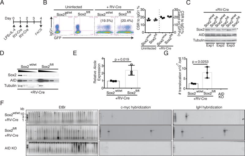
FIGURE 4.

Loss of Sox2 increases IgH:c-Myc translocations. (A) Strategy to delete Sox2 in ex vivo stimulated splenic B cells. B cells were harvested from wild type (Sox2wt/wt) or Sox2flox/flox (Sox2fl/fl) mice, activated with LPS+IL-4 and retrovirally infected with virus expressing Cre-recombinase and a GFP marker. (B) CSR to IgG1 was assayed by flow cytometry and quantified (n=3). (C) Cells were harvested and analyzed by immunoblotting for expression of Sox2, AID and tubulin (loading control). * represents a background polypeptide that would sometimes react with Sox2 antibodies. (D) Immunoblot with 2-fold dilution of protein extracts to assess AID levels. The 4 lanes for each genotype represent 10, 5, 2.5 and 1.25 μg of extract. (E) RNA from infected cells was analyzed for aicda levels by qPCR relative to β-actin; expression of wild type B cells was normalized to 1. (F) Genomic DNA from activated splenic B cells was subjected to two rounds of PCR, resolved on agarose gel, stained with Ethidium bromide (EtBr panel) and followed by Southern blot analysis with IgH and c-Myc probes to assess translocation frequency. (G) Translocation frequency; n=3.
