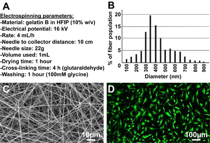
Figure 3. BIC characterization and biocompatibility.

(A) summarizes the electrospinning parameters used to produce the gelatin electrospun nanofibers depicted in (C), with an average fiber diameter of 350 nm (B). MIAMI cells seeded onto scaffolds remained viable and proliferated for at least 21 days, adopting an elongated morphology typical of MIAMI cells (D, confocal microscopy).
