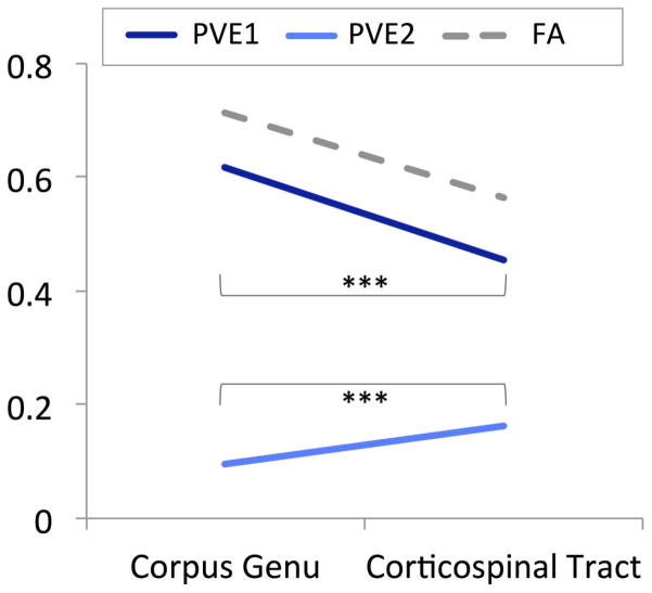
Figure 1. Partial volume estimates (PVEs) for primary and secondary fibers in the corpus genu and corticospinal tract.
Figure one shows partial volume estimates (PVEs) for a region of uniform fiber orientations (corpus genu) and for a region associated with crossing fibers (corticospinal tract). Across all participants (n=114), PVEs for primary fibers (PVE 1) were decreased and PVEs for secondary fibers (PVE 2) were increased in the corticospinal tract in comparison to the corpus genu. For reference, fractional anisotropy (FA) values are also shown. ***p<.001

