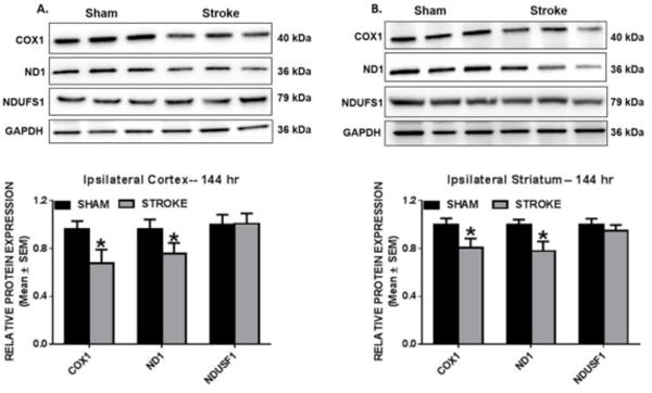
Fig. 4. Reduced mitochondrial encoded protein expression in the ipsilesional cortex and striatum.

Rats were subjected to either sham or ET-1 treatment. COX1, ND1, and NDUFS1 protein expression was determined by immunoblot analysis. These markers were measured in protein isolated from ipsilesional cortex (a) and striatum (b) 144hr following ET-1 exposure. GAPDH is used as loading control. Values reported as mean ± SEM. n =6–9, *p < 0.05
