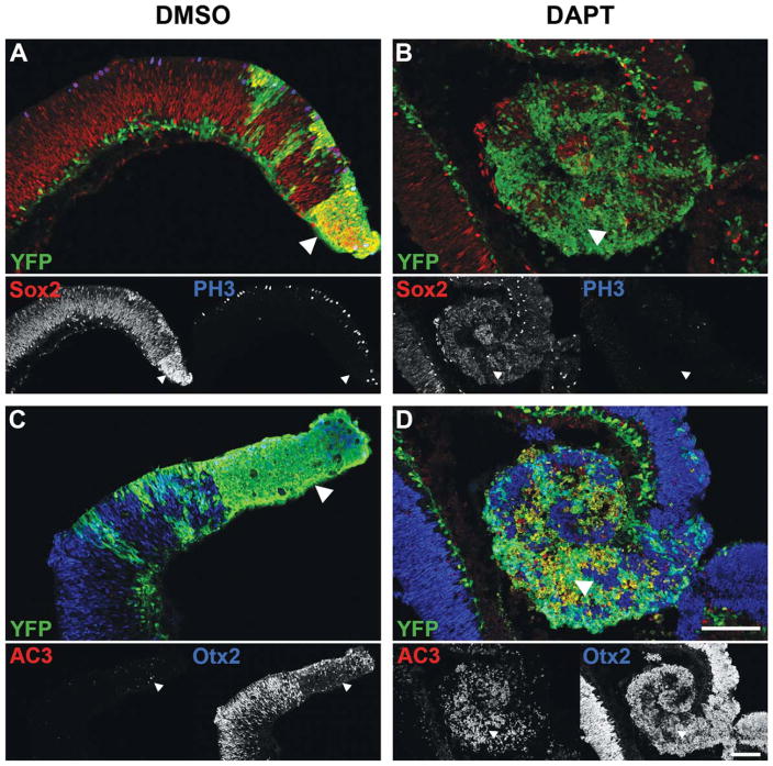
Figure 2.
Immunofluorescence staining of P0 Dicer CKO retinas after 48 h in culture. A–D, YFP staining (green, arrowheads) indicates areas of Dicer CKO. A,C, Immunofluorescence staining for the progenitor markers Sox2 (red) and PH3 (blue), as well as the apoptotic marker AC3 (red) and the photoreceptor marker Otx2 (blue) shows no change after 48h in DMSO from that described previously in vivo (Georgi and Reh, 2010). B,D, After 48 h in the Notch inhibitor DAPT, Dicer CKO cells (green, arrowheads) show a downregulation of Sox2 (red) and PH3 (blue), with a concomitant upregulation of Otx2 (blue), similar to that observed in wild type cells. Unlike wild type cells, Dicer CKO cells also show an induction of apoptosis, as indicated by increased AC3 staining (red). Scale bars: 100 μm. [Color figure can be viewed in the online issue, which is available at wileyonlinelibrary.com.]

