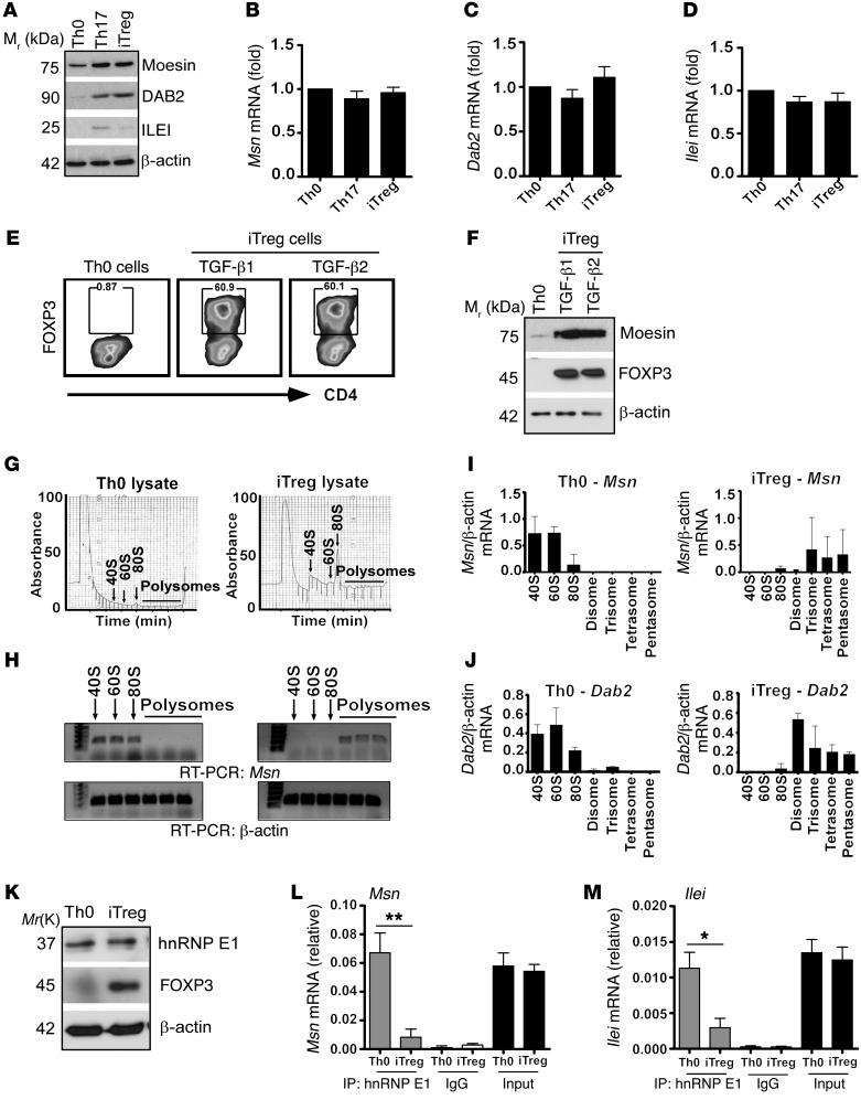
Figure 1. TGF-β translationally upregulates moesin mRNA expression within iTregs.
Primary CD4+CD25– T cells were isolated from C57BL/6 mice and stimulated with plate-bound anti-CD3 and soluble anti-CD28, IL-2, anti–IFN-γ, and anti–IL-4 without TGF-β for Th0 cells; or with IL-6 (50 ng/ml) and TGF-β (5 ng/ml) for Th17; and TGF-β1 (10 ng/ml) for iTregs for 3 days. (A) Immunoblotting of moesin, DAB2, and ILEI in T cells. (B–D) Fold mRNA of Msn (B), Dab2 (C), and Ilei (D) in Th0 and Th17 cells and iTregs. (E and F) Flow cytometry analysis (E) and immunoblot of FOXP3 expression (F) in iTregs using TGF-β1 or TGF-β2. (G and H) Polyribosome profiling (G) and RT-PCR (H) of Msn mRNA from monosomal fractions (40S, 60S, and 80S) in Th0 cells to the translating polysomal fractions in iTregs. (I and J) Quantitative RT-PCR of Msn (I) and Dab2 (J) mRNAs from the monosomal to polysomal fractions in iTregs. (K) Immunoblot of hnRNP E1, FOXP3, and β-actin in Th0 cells and iTregs. (L and M) RNA immunoprecipitation (RIP) of Msn and Ilei transcripts in Th0 cells and iTregs. Data represent the mean ± SD of 3 independent experiments performed in triplicate (B–D, I, and J), or 2 independent experiments performed in triplicate (L and M). *P < 0.05, **P < 0.01 by Student’s t test.

