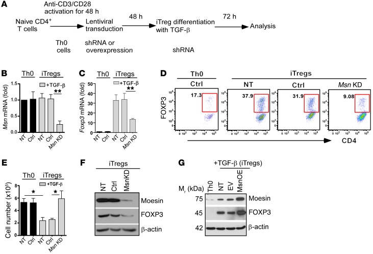
Figure 2. Moesin promotes FOXP3 induction in TGF-β–mediated iTregs.
(A) Lentiviral shRNA transduction steps to generate moesin knockdown (Msn KD) T cells. (B and C) Quantitative RT-PCR of total Msn (B) and Foxp3 (C) mRNA expression in Th0 cells and iTregs not transduced (NT) or transduced with control vector (Ctrl) or Msn shRNA. (D) Representative flow cytometry analysis of percentage FOXP3 expression in the transduced cells shown in B and C. (E) Assessment of cell numbers by trypan blue exclusion in iTreg differentiation culture. Data represent the mean ± SD of at least 3 independent experiments performed in triplicate (B, C, and E). *P < 0.05, **P < 0.01 by Student’s t test. (F) Immunoblot of moesin and FOXP3 in WT CD4+CD25– T cells first differentiated into iTregs in vitro, and then transduced with lentivirus shRNA or control. Representative data are shown. (G) Moesin overexpression (Msn OE) in primary CD4+CD25– T cells and FOXP3 expression in iTregs. EV, empty vector. Data shown are representative of at least 3 independent experiments.

