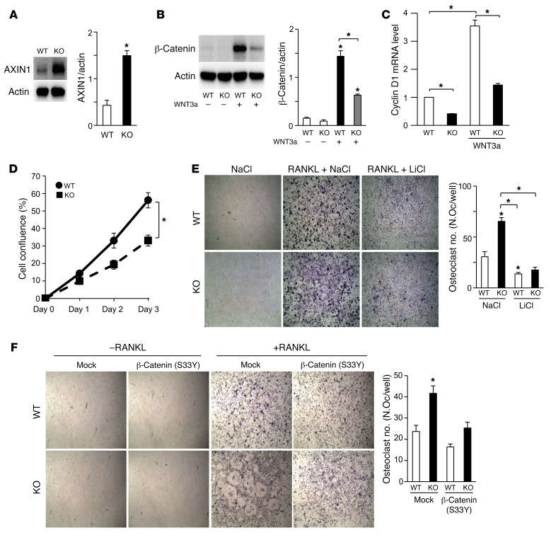
Figure 7. The Wnt/β-catenin pathway is impaired in Rnf146fl/fl LysM-Cre osteoclast progenitors.
(A) Whole cell lysates from primary murine osteoclast progenitors derived from WT and Rnf146fl/fl LysM-Cre (KO) mice were probed with the indicated antibodies for Western blot analysis. (B) Whole cell lysates from cells in A cultured in the presence or absence of a WNT3a-conditioned medium were probed with the indicated antibodies for Western blot analysis. (C) qPCR analysis of cyclin D1 mRNA expression in cells in B. n = 3. (D) Growth curves of primary murine osteoclast progenitors derived from WT and Rnf146fl/fl LysM-Cre (KO) mice cultured in a WNT3a-conditioned medium for 3 days. n = 3. (E) TRAP staining of osteoclasts derived from WT and Rnf146fl/fl LysM-Cre (KO) osteoclast progenitors cultured in the presence or absence of RANKL, NaCl (10 mM), and LiCl (10 mM) for 7 days. Original magnification, ×40. n = 3. (F) TRAP staining of osteoclasts derived from WT and Rnf146fl/fl LysM-Cre (KO) osteoclast progenitors infected with an empty vector control (mock) or β-catenin (S33Y)–expressing retroviral vector and cultured in the presence or absence of RANKL for 7 days. Original magnification, ×40. n = 3. P values were determined by ANOVA with Tukey-Kramer’s post hoc test (B, C, E, and F) or unpaired t test (A and D). Data are presented as mean ± SEM. *P < 0.05.

