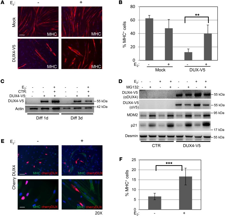
Figure 3. E2 antagonizes DUX4-mediated impairment of myoblast differentiation.
(A) Representative photographs of MHC immunostaining (red) in control myoblasts transfected with empty vector (mock, upper panels) or DUX4-V5 (lower panels) after 4 days of culture in differentiation medium in the absence or presence of E2. Nuclei were counterstained with DAPI (blue). Scale bar: 75 μm. (B) Percentage of MHC+ cells treated as in A. Mean ± SD of 2 independent experiments is shown. Three different fields for each condition were counted (n = 6). (C) Western blot of the indicated proteins in the lysates from myoblasts treated as in A. DUX4-V5 was detected by aV5 antibody. (D) Western blot of the indicated proteins in control myoblasts transfected as in A and collected after 72 hours of culture in differentiation medium with or without E2 and with 60 μM MG132 for the last 4 hours. See complete unedited blots for C and D in the supplemental material. (E) Representative photographs of MHC immunostaining (green) in control myoblasts transfected with Cherry-DUX4 (red) after 3 days of culture in differentiation medium in the absence or presence of E2. Nuclei were counterstained with DAPI (blue). Scale bars: 75 μm (upper panels); 25 μm (lower panels). (F) Quantification of double MHC+ Cherry-Dux+ cells. Mean ± SD of 2 independent experiments is shown. Four different fields for each condition were counted (n = 8). **P < 0.01; ***P < 0.001, 2-tailed Student’s t test.

