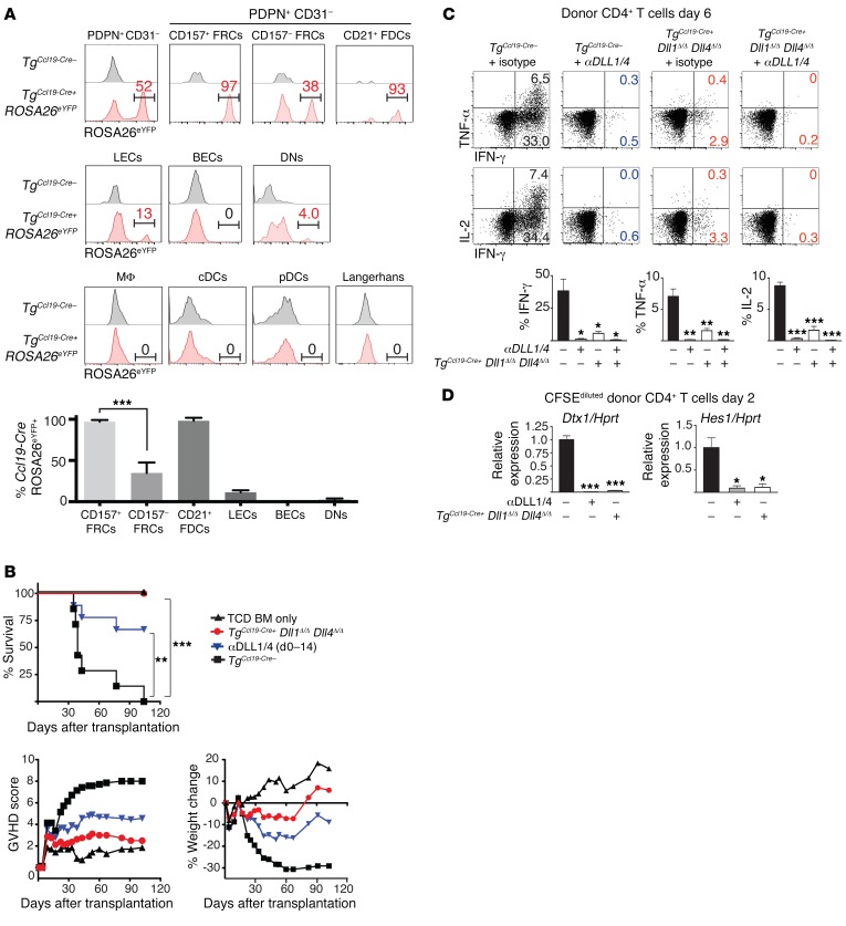
Figure 3. Ccl19-Cre+ lineage–traced stromal cells are the critical cellular source of DLL1/4 Notch ligands during acute GVHD.
(A) LNs were collected on day 1.5 after transplantation from lethally irradiated TgCcl19-Cre+ ROSA26eYFP mice receiving allogeneic BALB/c splenocytes and enzymatically digested. (A, top) eYFP expression in LN-resident bulk fibroblastic stromal cells (PDPN+CD31–) as well as subfractionated CD157+ FRCs, CD157– FRCs, and CD21+ FDCs. (A, middle) eYFP in LECs, BECs, PDPN–CD31– stromal cells (DNs). (A, bottom) eYFP in macrophages, conventional DCs (cDCs), pDCs, and skin-derived Langerhans cells. Bars in histograms define gating for eYFP+ cells, and numbers indicate the percentage of gated eYFP+ cells within parental cell populations, as identified by flow cytometric analysis. Bar graph in A shows the mean percentage of eYFP expression in each indicated nonhematopoietic subset (n = 4 mice/group; error bars indicate SD). (B) Survival, GVHD score, and weight of lethally irradiated (12 Gy) littermate control TgCcl19-Cre– or TgCcl19-Cre+ Dll1Δ/Δ Dll4Δ/Δ mice that were transplanted with 10 × 106 TCD BM only or 10 × 106 TCD BM plus 20 × 106 allogeneic BALB/c splenocytes. Isotype control or anti-DLL1/4–neutralizing antibodies were injected i.p. on days 0, 3, 7, and 10 (n = 10 mice/group). (C) Intracellular cytokines in donor CD4+ cells after anti-CD3/CD28 restimulation on day 6 (n = 5 mice/group). (D) Relative abundance of Dtx1 and Hes1 Notch target gene transcripts in donor CD4+ T cells sort purified from TgCcl19-Cre– plus isotype control, TgCcl19-Cre– plus anti-DLL1/4, or TgCcl19-Cre+ Dll1Δ/Δ Dll4Δ/Δ recipient mice on day 2 after transplantation (n = 5 mice/group). *P < 0.05, **P < 0.01, and ***P < 0.001, by unpaired, 2-tailed Student’s t test with Sidak’s correction for multiple comparisons. Data are representative of at least 5 experiments; error bars indicate SD.

