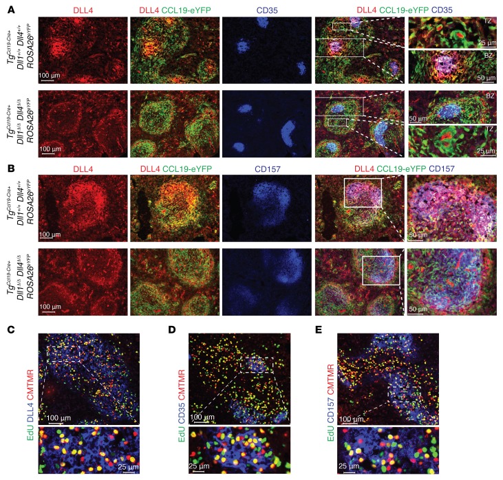
Figure 8. Fibroblastic niches in spleen express DLL1/4 Notch ligands and localize next to alloreactive T cells.
(A and B) Immunofluorescence microscopy of splenic cryosections from TgCcl19-Cre+ Dll1+/+ Dll4+/+ ROSA26eYFP and TgCcl19-Cre+ Dll1Δ/Δ Dll4Δ/Δ ROSA26eYFP mice stained for GFP, CD35, and DLL4 (A) or GFP, CD157, and DLL4 (B). (C–E) Immunofluorescence microscopy of splenic cryosections from lethally irradiated (8.5 Gy) BALB/c mice transplanted with CMTMR-labeled alloantigen-specific CD4+ 4C TCR–transgenic cells and pulsed with EdU 12 hours prior to organ collection to label proliferating cells. Cryosections were incubated with Alexa Fluor 488 picolyl azide to reveal EdU, along with anti-DLL4 (C), anti-CD35 (D), or anti-CD157 (E). Organs were collected on day 1.5 after transplantation. Data are representative of at least 2 experiments.

