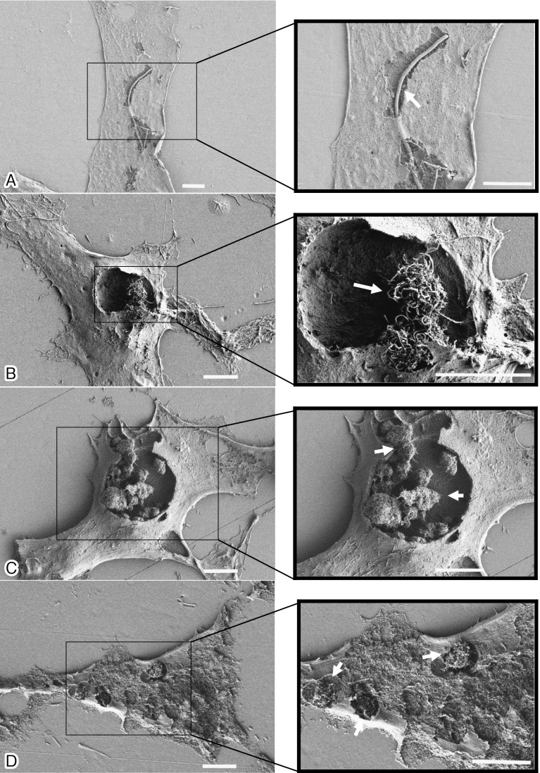
Fig. 2.
Four types of MWCNTs localized in the cytoplasm of CHL/IU cells can be observed in cells with partially removed surface membranes areas. The cells were exposed to the four MWCNTs at 2.5 (CTa) and 100 (CTd, CTe, and CTf) µg/ml for 48 h. A: An SEM image showing isolated long CTa fibers completely internalized into a CHL/IU cell. B: An SEM image showing entangled CTd fibers, similar to a bird’s nest, internalized into a cell. C: An SEM image showing a large CTe cluster completely internalized into a cell. D: An SEM image showing small CTf aggregates scattered in the cytoplasm of a cell. The white bar indicates 5 µm. Arrows indicate internalized MWCNTs.

