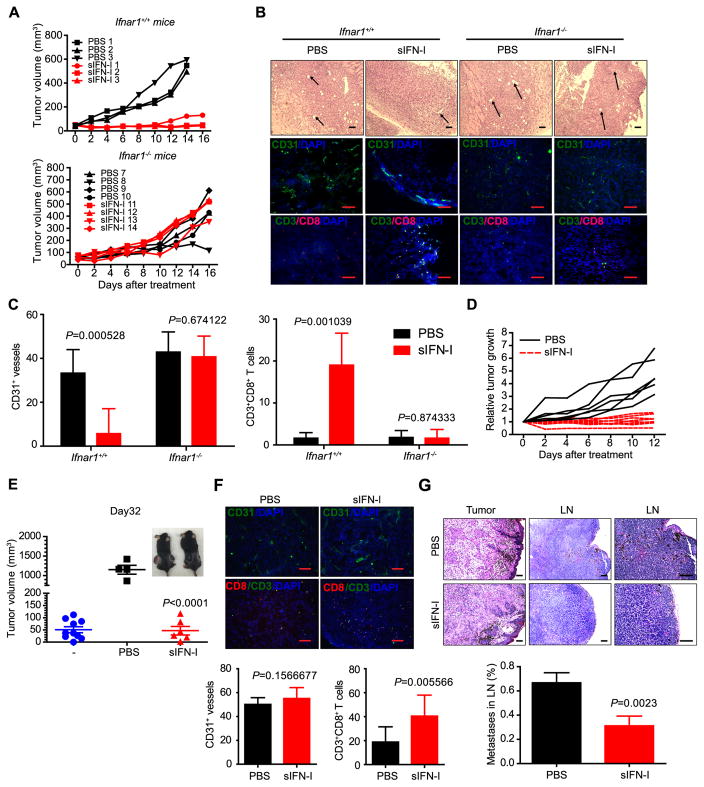
Figure 6. Anti-tumorigenic, anti-angiogenic and immunostimulating effects of sIFN-I in immunocompetent mouse models.
A. YUMM (BrafV600E/+; PtenΔ/Δ; CDKN2A−/−) cells were injected subcutaneously into Ifnar1+/+ and Ifnar1−/− mice to establish transplantable tumor model. sIFN-I or IFNα-2b (5 mg/kg) were injected intraperitoneally every other day for the indicated days. The tumor volume was measured and calculated.
B. H&E and immunofluorescence staining of YUMM tumors isolated from mice after sIFN-I treatment. Arrows indicate the vessels in tumor tissue. Scale bar, 100 μm.
C. Quantification on the positive CD31+ vessels number in the fields (n=7) and CD3+CD8+ T cells infiltrated in YUMM allograft tumor microenvironment (n=10).
D. Melanocyte-specific Cre activity was induced in adult mice (BrafCA/+Ptenf/f) by topical application of 4-HT to shaved back skin. Melanoma growth was measured after intraperitoneal injection with sIFN-I every other day.
E. Volume of melanoma tumors that grew in BrafV600E/+; PtenΔ/Δ mice to initial volume (“-”, blue circles). After that, mice were randomly assigned to two groups treated with PBS (black squares, left mouse in the inset) or sIFN-I (red triangles, right mouse at inset) for 32 days. p<0.001 between PBS and sIFN-I group.
F. Immunofluorescence staining of the tumor isolated from BrafV600E/+; PtenΔ/Δ mice after sIFN-I treatment. Bottom, quantification on the average positive CD31+ vessels number in the fields (n=10) and the double positive CD3+CD8+ cells in the fields (n=10) presenting infiltrated effector T cells in tumors from BrafV600E; PtenΔ/Δ mice. Scale bar: 100 μm.
G. H&E staining of the tumors and superficial lymph nodes (n=12) isolated from BrafV600E/+; PtenΔ/Δ mice after sIFN-I treatment. Bottom, quantification on the number of metastatic tumors in lymph node (LN). Scale bar, 100 μm.

