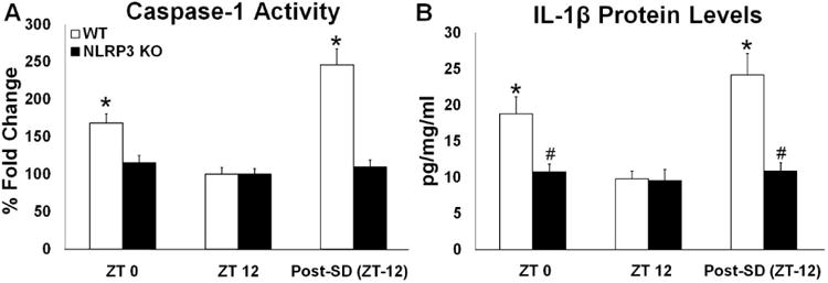
Fig 3.

Caspase-1 activity and IL-1β protein levels in the somatosensory cortex during light and dark periods during baseline sleep and post-SD (post-SD). WT but not NLRP3 KO mice exhibited significant enhancements in caspase-1 activity during the light period (ZT 0) compared to the dark period (ZT 12)(A). Caspase-1 activity post-SD was also significantly enhanced in WT but not NLRP3 KO mice when compared to time-of-day matched controls (ZT (12)(A). Similarly, IL-1β protein levels were significantly greater during the light period (ZT 0) compared to the dark period (ZT 12) and post-SD compared to time-of-day matched controls (ZT (12) only in WT control mice (B). N = 8 per genotype. (*) indicates significant difference between baseline and treatment. (#) indicates significant difference between genotypes. The Bonferroni corrected α value was set at p < 0.02.
