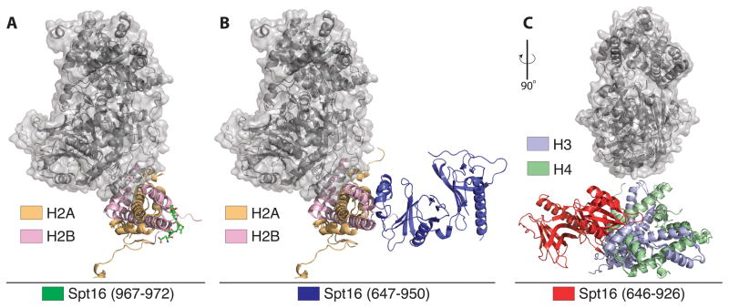
Figure 2.
Structural insights into relation of FACT and DUB module binding to histones. All panels show relative position of DUB module (gray surface) on either H2A/H2B or H3/H4. (A) Docking of DUB module relative to a peptide derived from the C-terminus of Spt16 solved to 1.80 Å resolution (PDB ID: 4WNN). (B) The Spt16M domain from Chaetomium thermophilum fused to H2B solved to 2.35 Å resolution (PDB ID: 4KHA). (C) Structure of the human FACT mid-AID domain bound to an H3/H4 tetramer solved to 2.98 Å resolution (PDB ID: 4Z2M).

