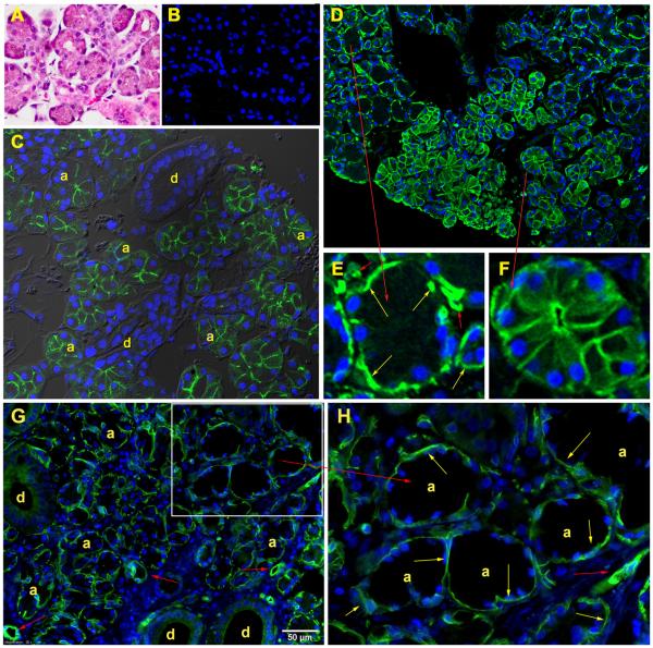
Figure 7.
Images from core needle biopsy specimen obtained from subject #19 at follow-up visit 1 (day 1124 after AdhAQP1 administration). A. H&E staining of the parotid gland tissue sample, showing the presence of acini and ducts. B. Control for immunofluorescence staining using normal rabbit IgG as the primary antibody. The nuclei are stained using DAPI and have a blue color. C. Tissue stained with an antibody to human AQP5. The immunofluorescence staining observed is localized only to the luminal membrane of acinar cells (a) and the closely adjacent intercalated duct region. Larger ducts (d) do not express AQP5 and are unstained. D. Tissue stained with an antibody to human AQP1. The immunofluorescence staining is found in three cell types: myoepithelial, vascular endothelial and acinar. Normally, AQP1 is only present in myoepithelial and vascular endothelial cells (Gresz et al 17). Acinar cells that can be seen expressing AQP1 (right central and bottom portion of panel) were transduced with AdhAQP1 administered to subject # 19 1124 days previously. E. An enlarged region of Panel D showing the presence of AQP1 in myoepithelial (yellow arrows) and vascular endothelial cells (red, smaller arrows) and the negative staining of a non-transduced acinus. F. An enlarged region of Panel D showing the abundant presence of AQP1 in the basolateral and luminal membranes of a transduced acinus. G. AQP1 localization in a biopsy specimen from a normal, male human volunteer’s parotid gland, i.e., without AdhAQP1 transduction. There is no immunofluorescence staining in acinar cells. H. An enlarged region of Panel G clearly showing the absence of AQP1 staining in acinar (a) and duct (d) cells, but its presence in myoepithelial cells (yellow arrows) and vascular endothelial cells (smaller red arrows). See SUBJECTS AND METHODS for details on the staining procedures and antibodies used.

