
Figure 1. MDM2 but not MDM4 maintains retinoblastoma cell proliferation.
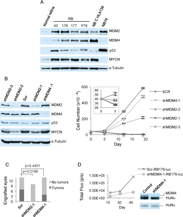
A. Western analysis of MDM2 (with SMP14), MDM4, p53, MYCN, and α-tubulin expression in post-fertilization week 19 human fetal retina and in four retinoblastoma (RB) and two neuroblastoma (NB) cell lines B. Western analysis at day 4 (left) and cell growth response (right) of RB176 cells after infection with lentivirus expressing shRNAs against MDM2 (shMDM2-1 and shMDM2-2) or against MDM4 (shMDM4-1), or expressing a scrambled shRNA control (Scr). Values and error bars denote mean and standard deviation (s.d.) of triplicate assays. C. Tumors formed up to 3.5 months after xenograft of RB176-luc cells transduced with shScr, shMDM2-1, or shMDM4-1 and engrafted into the sub-retinal space of athymic (nude) mice. P values are from two-tailed Fisher’s exact test. D. Representative tumor growth tracked over 1.5 months by bioluminescent imaging, (left) and MDM4 and HuNu expression in the tumors examined by Western blot sequentially probed with anti-HuNu (bottom) followed without stripping with anti-MDM2 (top).