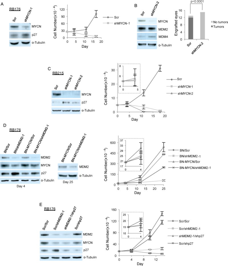
Figure 3. MDM2 maintains retinoblastoma cell proliferation in part by promoting MYCN expression.

A. Western analysis at day 4 (left) and cell growth response (right) of RB176 cells after infection with lentivirus expressing shRNA against MYCN or a scrambled control (Scr). B. Tumors formed by RB176-luc cells transduced with shScr but not by those transduced with shMYCN in mouse subretinal xenografts (right, P-value from two-tailed Fisher’s exact test). Western analysis of the cells used for xenograft (left). C. Western analysis at day 4 (left) and cell growth response (right) of CHLA-RB215 cells after infection with lentivirus expressing shRNA against MYCN or Scr. D. Western analysis at days 4 and 25 (left and middle) and cell growth response (right) of RB176 cells after co-infection with lentivirus expressing shRNA against MDM2 or Scr and with lentivirus expressing MYCN under control of the EF1α promoter (BN-MYCN) or the empty vector (BN). E. Western analysis at day 4 (left) and cell growth response (right) of RB176 cells after co-infection with lentivirus expressing shMDM2 or Scr and with lentivirus expressing shp27 or Scr.
