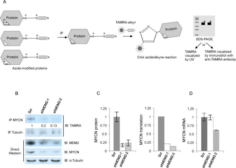
Figure 5. MDM2 regulates MYCN translation.

A. Diagram of protein translation assay. Nascent proteins were labeled with the azide-modified alanine analog- azidohomoalanine (AHA), immunoprecipitated, and incorporated AHA reacted with a fluorescent TAMRA labeled alkyne. TAMRA labeled proteins were separated by SDS-PAGE and detected by immunoblot using anti-TAMRA antibody (in Fig. 5b) or by UV fluorescence (in Supplementary Fig. S4). B. Protein translation assay performed on AHA-labeled RB176 cells 4 days after infection with lentivirus expressing either of two shRNAs against MDM2 or a scrambled control (Scr). AHA-labeled cell lysates either were sequentially immunoprecipitated with MYCN and then α-tubulin antibodies followed by anti-TAMRA immunoblot (top) or were used for direct Western analysis of MDM2, MYCN, and α-tubulin (bottom). C. Quantitation of total MYCN protein (the average and s.d. of two western analyses of the same lysate) and newly translated MYCN protein, both normalized to α-tubulin. D. qRT-PCR analysis of MYCN RNA from the same AHA-labeled cells as used for protein analyses (average and s.d. of triplicate technical replicates).
