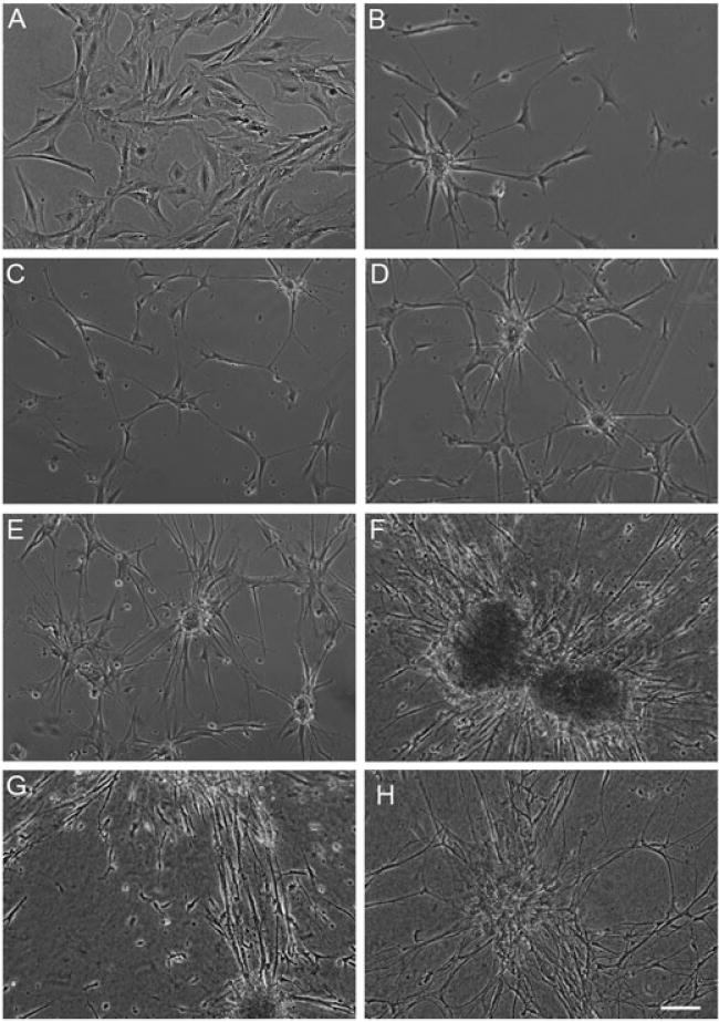
Figure 2.

Optimization of culture conditions resulted in astrocytic clustering and numerous processes. Brightfield microscopy images of astrocytes cultured on polystyrene in (A) serum-containing medium (DMEM/F12 + 10% FBS) vs (B) defined, serum-free medium (neurobasal +2% B27 + 1% G-5 + 0.25% L-glutamine) produced astrocytes with a mature, non-proliferative, process-bearing morphology. Astrocytes cultured for 5 days in vitro (substrates precoated with poly-L-lysine), grown on (C) polystyrene only, (D) ACLAR only, (E) 1 mg/ml Matrigel-coated ACLAR and (F) 1 mg/ml rat tail collagen type I-coated ACLAR. (C–F) Cultures were plated with de-fined media and astrocytes cultured on rat tail collagen type I showed the most robust clustering and processes at both (G) 5 and (H) 20 days in vitro; scale bar =100 μm
