
Figure 4.
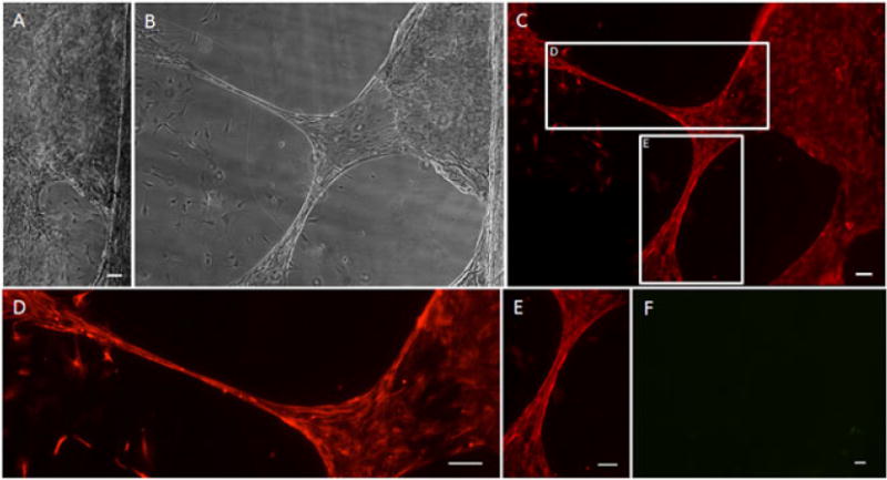
Stretched astrocyte processes express glial fibrillary acidic protein (GFAP). After the application of continuous mechanical tension at a rate of 0.3 mm/day (12.5 μm/h), astrocyte processes were successfully ‘stretch-grown’. (A) Brightfield image of culture prior to application of continuous mechanical tension; scale bar =50 μm. (B) Brightfield image of the same area from (A) after stretch. (C) Fluorescence image of the same area, showing labelling for GFAP in red, confirming the astrocytic phenotype of the stretch-grown cells and processes; (D, E) enlargements of the stretched regions from (C). (F) Fluorescence of the same area from (B), showing absence of labelling for β-tubulin III in green, demonstrating that there were no contaminating neurons/neurites in the culture; scale bar (C–F) =100 μm