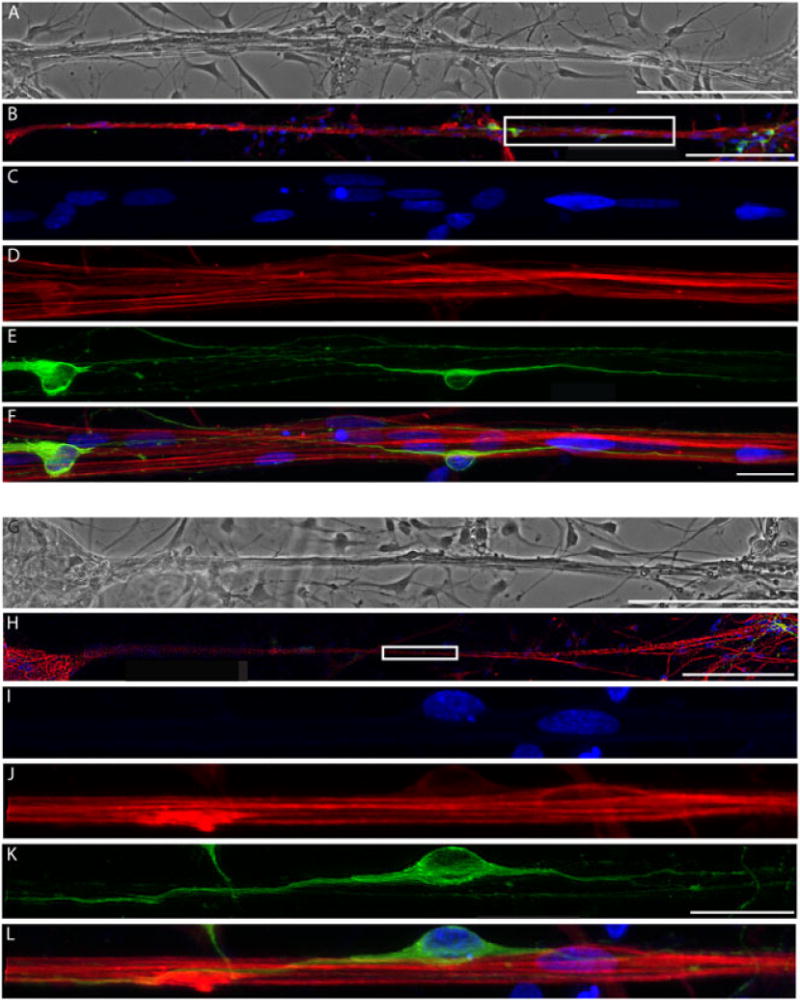
Figure 9.

Neurons extend neurites directly along the stretched astrocytic processes. (A, G) Phase-contrast image and (B, H) confocal reconstruction of two separate areas of astrocytic processes that were stretched to unprecedented lengths of 1.5 mm and directed and supported neuronal survival; scale bars =200 μm. (C–F) Enlargement of boxed region in (B), showing (C) Hoechst counterstain, (D) GFAP-positive stretched astrocytic processes, (E) β-tubulin III-positive neurons and (F) overlay; scale bars = 25 μm. (H) Second area demonstrating stretched astrocytic bundles seeded with neurons were (I) stained for Hoechst, (J) labelled for GFAP, (K) β-tubulin III (Tuj1) and (L) overlay
