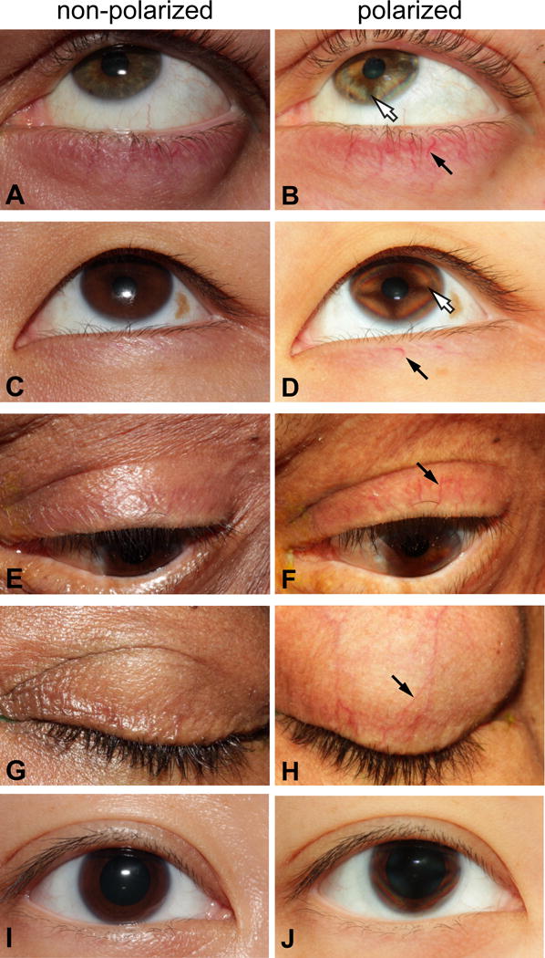
Figure 2.

Non-polarized and cross-polarized eyelid photographs. Conventional, non-polarized images of the eyelids of patients with blepharitis (panels A, C, E, and G) and the corresponding images of the same eyes through crossed-polarizers (panels B, D, F, and H). The method is illustrated in patients with blepharitis of different races (A, B: Caucasian; C, D: South Asian; E, F: lightly pigmented African-American; G, H: more heavily pigmented African-American). The control subject (I and J) is Asian. Black arrows indicate a representative blood vessel in each patient that is made more visible with cross-polarizers. One patient (E,F) has a loose eyelash visible on the upper eyelid (not highlighted). White arrows in B, D indicate the typical isogyre when the cornea is visualized through crossed-polarizers. No adjustments were made to any of the images other than the insertion of arrows and labeling.
