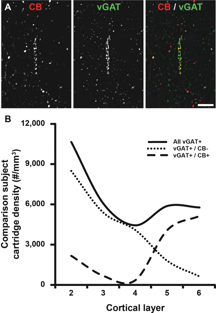
Figure 3.
ChC vGAT+/CB+ cartridges in human PFC. (A) Projection image (3 z-planes separated by 0.25 µm) of a human PFC tissue section immunolabeled for CB and vGAT. Single (CB and vGAT) and merged immunoreactive channels shown. (B) The density of all vGAT+ cartridges regardless of CB immunoreactivity (solid line), vGAT+/ CB negative cartridges (CB−; dotted line), and vGAT+/CB+ cartridges (dashed line) in cortical layers 2–6 of the human PFC.

