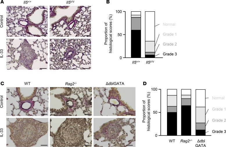
Figure 3. Advanced medial hypertrophy and progressive intimal proliferation of pulmonary arteries, which are dependent on eosinophils in IL-33–treated mice.
(A) Elastica van Gieson (EVG) staining of representative sections from Il5+/+ or Il5V/V mice, treated with PBS (control) or IL-33 on days 0, 7, and 14 (n = 4 per group). (B) Histological scoring of arterial hypertrophy. Seventy arteries were randomly selected from IL-33–treated Il5+/+ or Il5V/V mice (n = 4 per group) and scored according to arterial conditions (Supplemental Figure 4). (C) EVG staining of representative sections from WT, Rag2-deficient, and ΔdblGATA mice treated with PBS or IL-33 on days 0, 7, and 14 (n = 5 for WT and ΔdblGATA mice, and n = 6 for Rag2-deficient mice). (D) Samples of 84, 64, or 72 randomly selected arteries from WT, Rag2-deficient, or ΔdblGATA mice, respectively, were scored. Scale bars: 50 μm (A and C).

