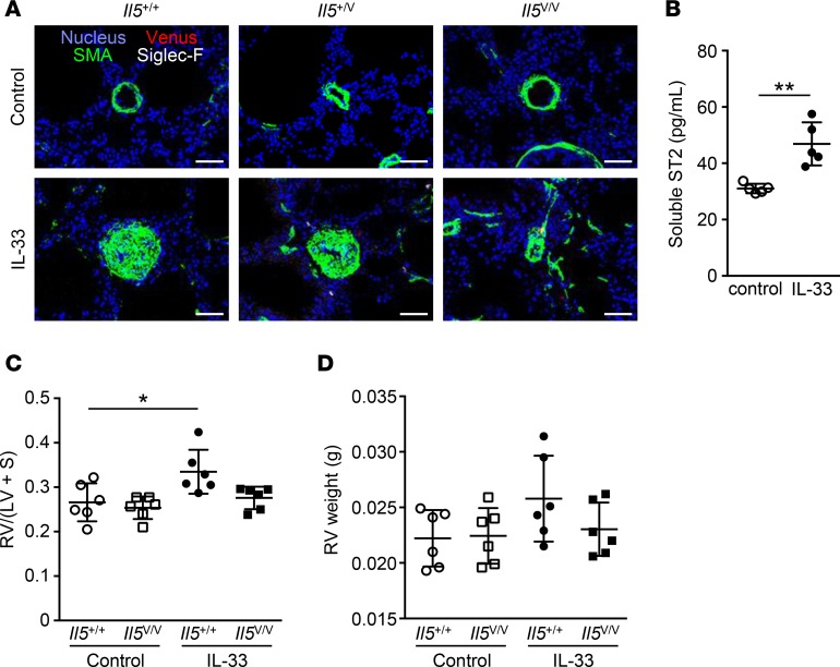
Figure 6. Prolonged chronic exposure to IL-33 resulted in disappearance of perivascular ILC2s and eosinophils, increased levels of serum soluble ST2, and right ventricular hypertrophy.
(A) Immunofluorescent histology of lungs from Il5+/+, Il5+/V, or Il5V/V mice treated with 11 weekly injections of PBS (control) or IL-33 (n = 4 per group). Venus+ cells (red) are shown with Siglec-F+ eosinophils (white) and smooth muscle actin+ (SMA) smooth muscle cells (green). The nuclei are visualized in blue. Scale bars: 50 μm. (B) Serum soluble ST2 from mice treated with PBS or IL-33 at 11 weekly intervals was measured by ELISA (n = 5 per group). P values were calculated using the two-tailed Student’s t test. (C and D) Comparison of right ventricular hypertrophy between Il5+/+ or Il5V/V mice treated with 11 weekly injections of PBS or IL-33 (n = 6). The right ventricular free wall (RV) and the left ventricle plus septum (LV + S) were weighted, and the degree of hypertrophy is shown as RV/(LV + S) ratios (C) and the absolute RV weight (D). Graph data are shown as means ± SD. P values were calculated using one-way ANOVA with Bonferroni test. Asterisks indicate statistical significance (*P < 0.05; **P < 0.01).

