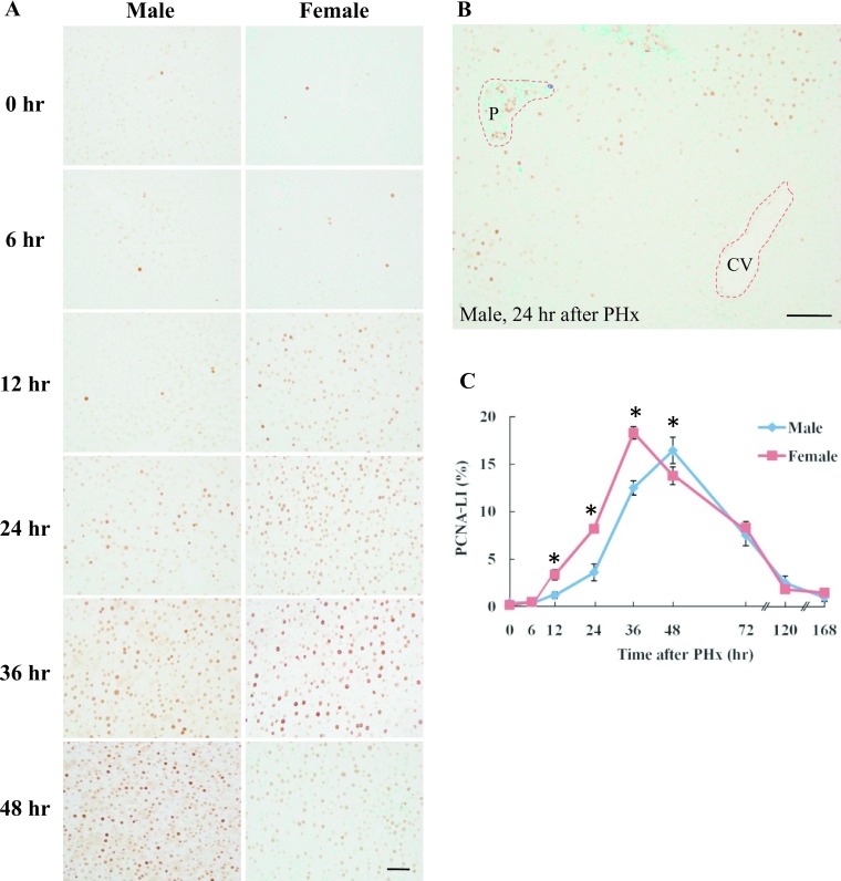
Fig. 2.
Immunohistochemical detection of PCNA in male and female rat liver after PHx. A: Liver tissue was collected from male and female rats at 0, 6, 12, 24, 36 and 48 hr after PHx. Paraffin-embedded rat liver sections were analyzed by immunohistochemistry. Magnification ×400. Bar = 50 μm. B: Zonal distribution of PCNA-positive cells at 24 hr in male rat liver after PHx. P: portal area, CV: central vein. Magnification ×200. Bar = 100 μm. C: PCNA-LI in male and female rats after PHx. The number of PCNA positive hepatocytes was counted at each time-point after PHx. Blue and red lines represent male and female, respectively. Asterisks indicate statistically significant differences (*p < 0.05). Data represent the mean ± SE of three independent experiments.

