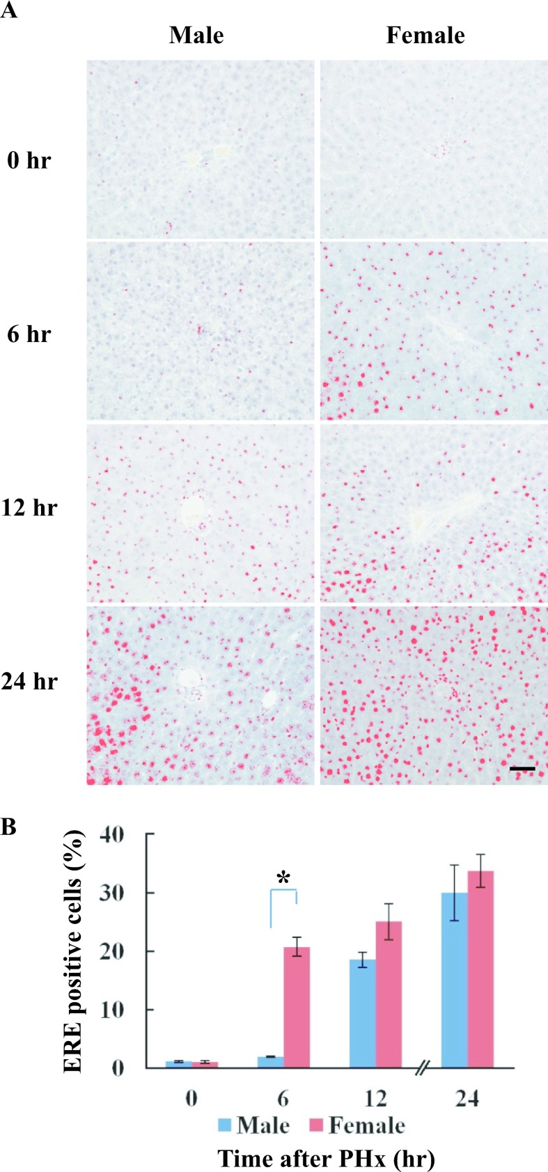
Fig. 4.
Localization of ERE binding proteins in male and female rats after PHx. Paraffin-embedded liver sections were used for detection of ERE binding proteins by Southwestern histochemistry at various time-points after PHx. A: The localization of ERE binding proteins was detected from 12 hr after PHx in male rats, but was found from 6 hr in female rats. Positive staining was processed using a DAB-image analyzer. B: The number of ERE-positive cells in the liver sections of male and female rats at various time-points after PHx. Blue and red columns represent male and female, respectively. Asterisk indicates statistically significant difference (*p < 0.05). Magnification ×400. Bar = 50 μm.

