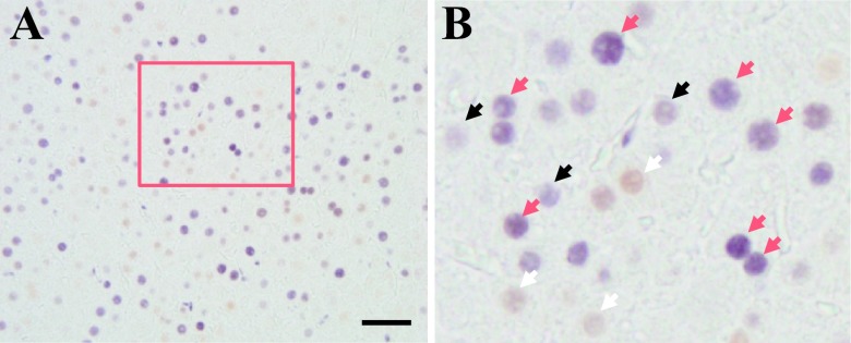
Fig. 5.
Double staining for PCNA and ERα in male and female rat liver after PHx. The paraffin-embedded sections were analyzed by immunohistochemistry. A: ERα-positive cells were stained brown (DAB), whereas PCNA positive cells were stained purple-blue (4-Cl-1-Naphtol). Boxed area is enlarged in B. Arrows indicate white (ERα), black (PCNA) and red (double staining), respectively. Magnification ×400. Scale Bar = 50 μm.

