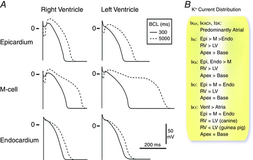
Figure 1. Heterogeneity of canine ventricular repolarization and K+ current distribution.

A, epicardial, M‐cell and endocardial APs from the RV and LV of the canine heart at basic cycle lengths (BCLs) of 300 and 5000 ms. Redrawn with permission from Antzelevitch et al. (1999). B, relative distribution of major K+ currents in the heart (see text for details and references).
