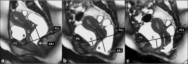
Fig. 3.
Basic measurements. a. Dynamic Balanced Fast Field Echo (BFFE) sequence in the midsagittal plane at rest shows how to plot the basic measurements of pelvic organ prolapse. The pubococcygeal line (PCL), drawn on sagittal plane from the inferior aspect of the pubic symphysis (PS) to the last coccygeal joint. After defining the PCL, the distance from each reference point is measured perpendicularly to the PCL at rest and at maximum straining. B; bladder base, C; cervix, P; pouch of Douglas, ARJ; Anorectal junction. Measured values above the reference line have a minus sign, values below a plus sign. b. Dynamic BFFE during maximum straining shows the movement of the organs compared to their location at rest. It is recommend to give the difference of the values at rest and during straining for each organ-specific reference point (pelvic organ mobility). R; Rectocele, ARJ; Ano-Rectal Junction. c. MRI defecography (BFFE) in the mid sagittal plane during evacuation of the intra-rectal gel. Dynamic MR imaging during evacuation is mandatory, because certain abnormalities and the full extent of POP are only visible during evacuation. In this case compared to the maximum staining phase it is obvious that there is increase of the degree of the pelvic organ descent and development of new pathology including the loss of urine and the detection of masked intussusception, which was detected only during excavation (white arrow)

