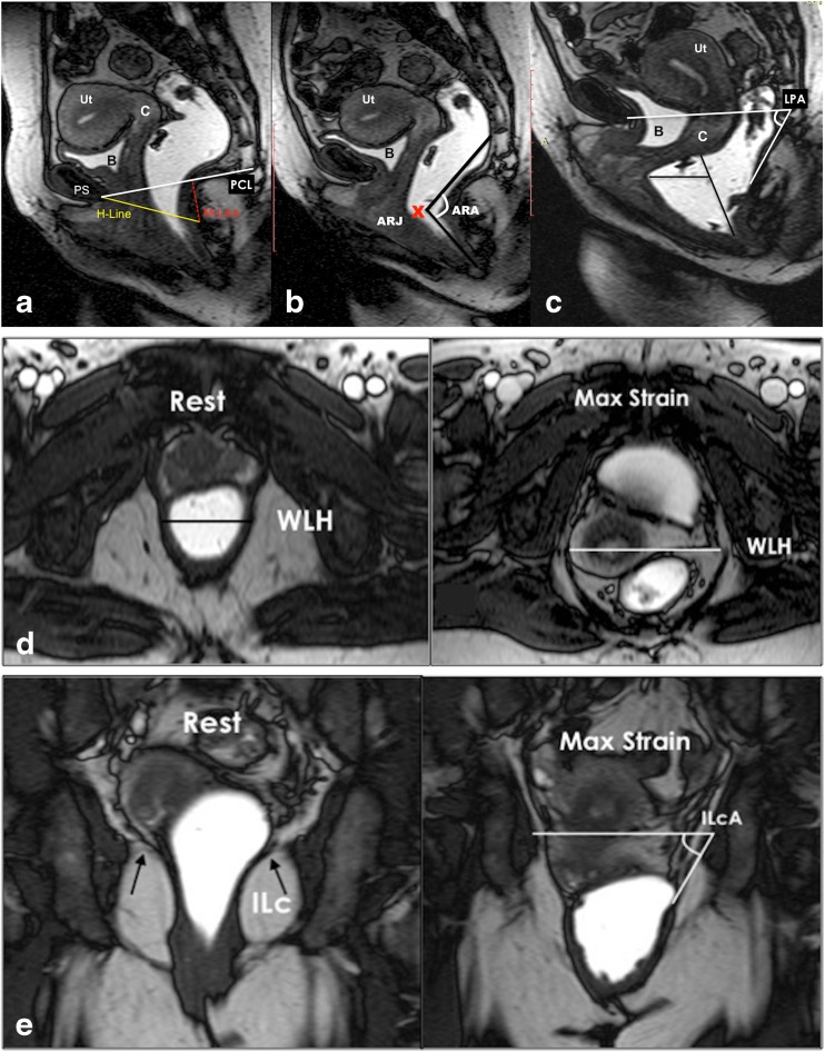
Fig. 4.
Pelvic floor relaxation and posterior compartment measurements. a,b,c Dynamic Balanced Fast Field Echo (BFFE) sequence in the midsagittal plane at rest (a) , mild (b), and maximum straining (c). (a) shows how to quantify the pelvic floor laxity. The H-line extends from the inferior aspect of the pubic symphysis to the anorectal junction, the M-line is dropped as a perpendicular line from the pubococcygeal line (PCL) to the posterior aspect of the H-line. (b) Demonstrates the anorectal angle (ARA) drawn along the posterior border of the rectum and a line along the central axis of the anal canal on sagittal plane. ARJ; Ano-Rectal Junction. (c) Shows how to measure and diagnose a pathological rectocele: a line drawn through the anterior wall of the anal canal is extended upward, and a rectal bulge of greater than 2 cm anterior to this line is described as a rectocele (R). The levator plate angle (LPA) is enclosed between the levator plate and the PCL. d,e. Dynamic Balanced Fast Field Echo (BFFE) sequence in axial (d) and coronal (e) plane at rest and during maximum straining. In the axial plane the width of the levator hiatus is enclosed between the puborectalis muscle slings. On the coronal plane, the iliococcygeus angle is measured between the iliococcygeus muscle and the transverse plane of the pelvis in posterior coronal images at the level of the anal canal

