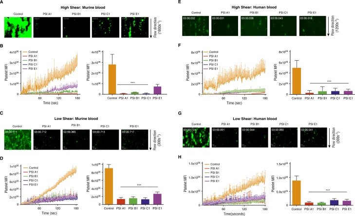
Figure 6.
Anti-β3 PSI mAbs inhibited thrombus formation in ex vivo perfusion chambers. Antibodies against PSI domain (10 μg/mL) were incubated with fluorescently labeled human or murine platelets and perfused over a collagen-coated surface (100 μg/mL). (A) Representative images at 3 minutes of murine platelets that were untreated (Control) or incubated with anti-PSI mAbs (PSI A1, PSI B1, PSI C1, and PSI E1) (high shear: 1800s−1). (B) Pretreatment with anti-PSI mAbs inhibited murine platelet adhesion and thrombus formation at a shear rate of 1800s−1 (equivalent to flow in stenotic vessels) (mean ± SEM; ***P < .001 [at 3 min]; n = 3 each). (C) Representative images at 3 minutes of murine platelets that were untreated (Control) or incubated with anti-PSI mAbs (PSI A1, PSI B1, PSI C1, and PSI E1) (low shear: 300s−1). (D) Pre-treatment with anti-PSI mAbs inhibited murine platelet adhesion and thrombus formation at a shear rate of 300s−1 (mean ± SEM; ***P < .001 [at 3 min]; n = 3 each). (E) Representative images at 3 minutes of human platelets that were untreated (control) or incubated with anti-PSI mAbs (PSI A1, PSI B1, PSI C1, and PSI E1) (high shear: 1200s−1). (F) Pretreatment with anti-PSI mAbs inhibited human platelet adhesion and thrombus formation at a shear rate of 1200s−1 (equivalent to flow in stenotic vessels) (mean ± SEM; ***P < .001 [at 3 min]; n = 3 each). (G) Representative images at 3 minutes of human platelets that were untreated (Control) or incubated with anti-PSI mAbs (PSI A1, PSI B1, PSI C1, and PSI E1) (low shear: 300s−1). (H) Pretreatment with anti-PSI mAbs inhibited human platelet adhesion and thrombus formation at a shear rate of 300s−1 (mean ± SEM; ***P < .001 [at 3 min]; n = 3 each).

