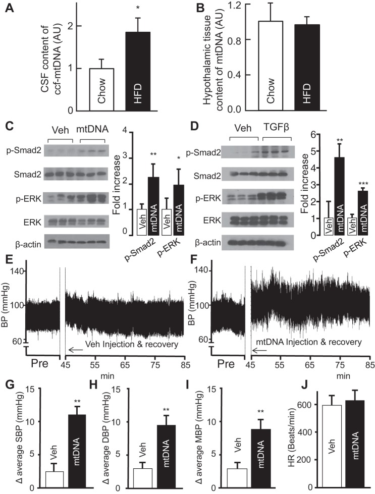
Fig. 1.
Obesity-associated brain excess in circulating mtDNA and the effect on blood pressure (BP). A and B: C57BL/6 mice fed on a HFD vs. normal chow for 12 wk were killed to obtain cerebrospinal fluid and hypothalamic tissues. Data show the fold increases of circulating cell-free (ccf) mtDNA in the cerebrospinal fluid (A) and tissue mtDNA in the hypothalamus (B) of HFD-fed mice compared with chow-fed mice. AU, arbitrary unit. C and D: standard C57BL/6 mice were preimplanted with an injection cannula in the hypothalamic third ventricle. After ~1-wk postsurgery recovery, they received a single injection of mtDNA (60 pg) or TGFβ1 (4 ng) in the third ventricle. Vehicle (Veh)-treated mice were used for comparison in each experiment. Western blots from mtDNA (C) or TGFβ1 (D)-treated mice were used to measure the levels of phospho-Smad2 (p-Smad2) and phospho-ERK (p-ERK) and quantified in graphs at right. E–J: regular C57BL/6 were preimplanted with a telemetric transmitter in the carotid artery and an injection cannula in the hypothalamic third ventricle. All mice were monitored for BP and heart rate following 1-wk postsurgery recovery. E and F: representative preinjection (Pre) period and 40-min telemetric BP tracings during the rest phase of a day. The time of 0–45 min was used for injection and postinjection recovery. G–J: average levels of systolic BP (G), diastolic BP (H), mean BP (I), and heart rate (J). SBP, systolic BP; DBP, diastolic BP; MBP, mean BP; HR, heart rate. *P < 0.05, **P < 0.01, ***P < 0.001; n = 3–6 mice per group (A–D) and n = 6 mice per group (G–J). Error bars reflect means ± SE.

