Fig. 2.

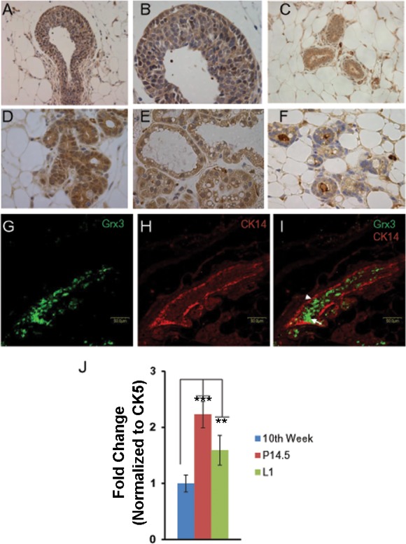
Grx3 expression at different mammary gland developing stages. A and B: terminal end bud of 7-wk-old virgin mice; Grx3 proteins were detected in cap, myoepithelial, luminal epithelial, most of body cells, and stroma cells. Grx3 proteins were examined in myoepithelial cells and luminal epithelial cells in gland ducts at 10th week of age in virgin mice (C), at pregnancy day 14 (D), at lactation day 1 (E), at involution day 7 (F). 200× magnification for A, 400× magnification for B–F. G–I: double immunolabeling of mammary gland tissue sectioned from MG at P14.5 with antibodies against Grx3 (G) and CK14 (H). I: shown is the merged image with Grx3 in luminal epithelial cells (arrow) and CK14 is present in myoepithelial cells (arrowhead). Scale bars, 50 µm. J: q-PCR analysis of Grx3 mRNA levels in mammary epithelial cells (MECs) isolated from glands at 10th week, pregnancy day 14.5, and lactation day 1. Cytokeratin 5 (CK5) was used as an internal control. Student’s t-test; n = 3. **P < 0.01, L1 vs. 10th week; ***P < 0.001, P14.5 vs. 10th week.
