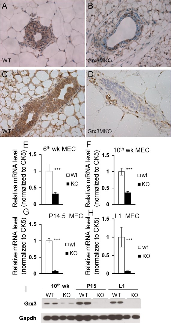
Fig. 3.

Grx3 deletion in MMTV-Cre/ Grx3flox/flox mammary glands. A–D: immunohistochemical staining at 6th week of age in virgin mice (A and B) and at pregnancy day 14 mice (C and D) shows Grx3 expression in ducts of floxed (A and C) and knockout (B and D) MGs. 400× magnification. E–H: q-PCR analysis of Grx3 expression in MECs from 6th week (E), 10th week (F), pregnancy day 14.5 (G), and lactation day 1 (H). Student’s t-test; n = 3. ***P < 0.001, significant difference between WT and KO. Cytokeratin 5 (CK5) was used as an internal control. I: Western blotting analysis of Grx3 protein levels in WT and KO MECs isolated from 10th week, P15, and L1 of MGs.
