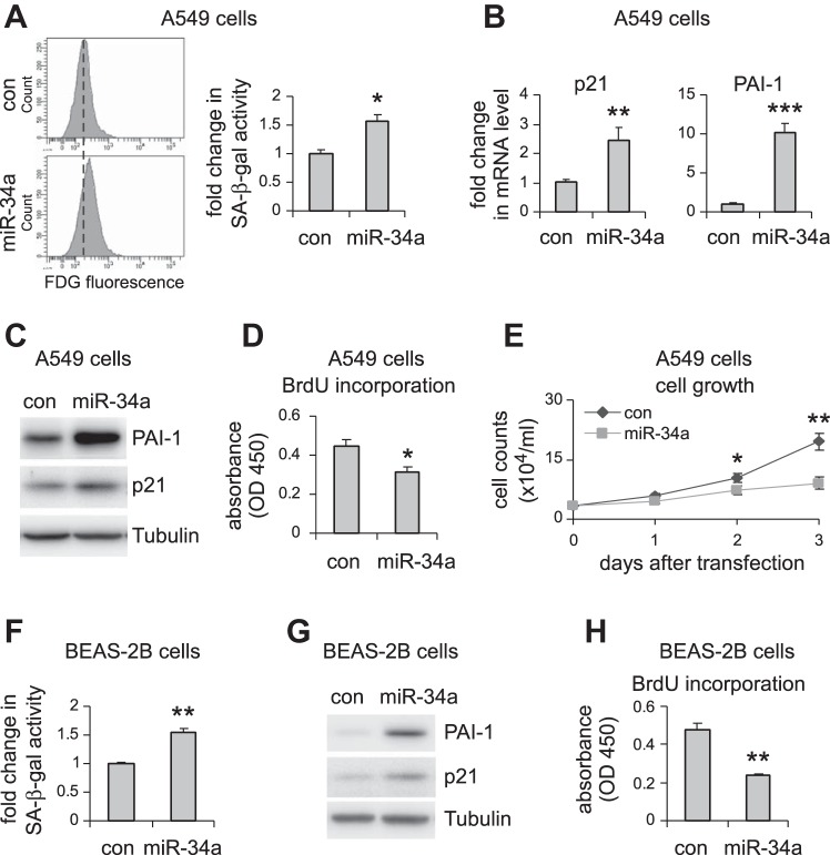
Fig. 3.
miR-34a induces cellular senescence and inhibits cell proliferation in pulmonary epithelial cells in vitro. A–D: human pulmonary epithelial cells A549 were transfected with 50 nM control (con) mimics or miR-34a mimics. A: 3 days after transfection, cells were collected, and senescence-associated β-galactosidase (SA-β-gal) activities in the cells were assessed by flow cytometry. mRNA and protein levels of p21 and PAI-1 were determined by real-time PCR (B) and Western blotting (C). D: cell proliferation was assessed by BrdU incorporation assay. E: A549 cells were transfected with con mimics or miR-34a mimics. Cells were trypsinized and counted at the indicated time points after transfection. A, B, D, and E: values are means ± SD; n = 3. *P < 0.05, **P < 0.01, and ***P < 0.001 compared with con miR group. F–H: human lung epithelial cells BEAS-2B were transfected with con mimics or miR-34a mimics as in A–D. Three days after transfection, cells were collected, and SA-β-gal activities in the cells were assessed by flow cytometry (F), PAI-1 and p21 levels by Western blotting (G), and cell proliferation by BrdU incorporation assay (H). F and H: values are means ± SD, n = 3. **P < 0.01 compared with con miR group. FDG, fluorescein di-β-d-galactopyranoside; OD, optical density.

